Diaphragm fragment detection
Diaphragm rupture is integral to the operation of shock and expansion tubes. A diaphragm initially separates two regions of gas at different pressures. A shock wave propagating down the tube causes the diaphragm to rupture by impulsively subjecting it to a large pressure differential. Although the influence of the diaphragm on the test flows generated in these facilities has long been recognised, the rupture process has not been understood. The aim of this work was to optically characterise the rupture process of a light diaphragm made of cellophane, as typically used in an expansion tube.

The experimental method was based on absorption of light by cellophane, and refraction of light due to changes in gas density and species. A series of holographic images was recorded at various times during the process of rupture of a diaphragm mounted at the interface between carbon dioxide test gas and low density helium accelerator gas.
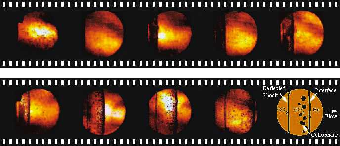
Clearly visualised in the experimental results, shown in the adjacent movie strip, is the evolving behaviour of the diaphragm material and gas discontinuities throughout the rupture process. Initially, the diaphragm bulges out into the lower pressure accelerator gas. After impact by a shock wave, the diaphragm breaks around its periphery and flattens. The diaphragm begins to fragment in the centre. As it moves downstream, it continues to disintegrate and some pieces of diaphragm material lag behind the propagating gas interface. A shock formed by reflection off the diaphragm is gradually swept downstream. Fragmentation of the cellophane in the field of view is complete by the time the last image was recorded, about 20 microseconds after shock impact: the diaphragm no longer blocks the flow.
Trajectories of the interface and reflected shock have been constructed using measurements from the holographic images. These imply that the diaphragm and reflected shock are subjected to accelerations that are initially very large but rapidly decrease. The diaphragm inertia model proposes that the diaphragm experiences decreasing pressure, and hence acceleration, due to motion down the tube as a single piece, like a piston. The images show that, for about 10 microseconds after shock impact, the physical system matches this description. During this period the trajectory predicted by the diaphragm inertia model provides a good fit to the experimental data. However, at later times the interface accelerates more than predicted. This may be explained by the effective reduction in mass of the diaphragm as it fractures and fragments fall behind. Therefore the central assumption of the diaphragm inertia model is invalidated at that stage.